ADCAIJ: Advances in Distributed Computing and Artificial Intelligence Journal
Regular Issue, Vol. 13 (2024), e31528
eISSN: 2255-2863
DOI: https://doi.org/10.14201/adcaij.31528
A Review on Covid-19 Detection Using Artificial Intelligence from Chest CT Scan Slices
Dhanshri Malia and S. A. Patilb
a Department of Electronics Engineering, DKTE Ichalkarnji Research Center, Shivaji Univesity, Kolhapur
b Head of Department Electronics Engineering, DKTE Ichalkarnji, Shivaji Univesity, Kolhapur
malidhanshri93@gmail.com, sapatil@dkte.ac.in
ABSTRACT
The outbreak of COVID-19, a contagious respiratory disease, has had a significant impact on people worldwide. To prevent its spread, there is an urgent need for an easily accessible, fast, and cost-effective diagnostic solution. According to studies, COVID-19 is frequently accompanied by coughing. Therefore, the identification and classification of cough sounds can be a promising method for rapidly and efficiently diagnosing the disease. The COVID-19 epidemic has resulted in a worldwide health crisis, and stopping the disease's spread depends on a quick and precise disease diagnosis. COVID-19 has been detected using medical imaging modalities such as chest X-rays and computed tomography (CT) scans due to their non-invasive nature and accessibility. This research provides an in-depth examination of deep learning-based strategies for recognising COVID-19 in medical images. The benefits and drawbacks of various deep learning approaches and their applications in COVID-19 detection are discussed. The study also examines publicly available datasets and benchmarks for evaluating deep learning model performance. Furthermore, the limitations and future research prospects for using deep learning in COVID-19 detection are discussed. This survey's goal is to offer a comprehensive overview of the current state of advancement in deep learning-based COVID-19 detection using medical images. This can aid researchers and healthcare professionals in selecting appropriate approaches for an effective diagnosis of the disease.
KEYWORDS
artificial intelligence; Coronavirus; COVID-19; deep learning and machine learning
1. Introduction
The SARS-CoV-2 virus, which is the source of the highly infectious respiratory ailment known as COVID-19, first surfaced in Wuhan, China, in December 2019 and quickly spread around the world, causing a global pandemic. COVID-19 primarily spreads through respiratory droplets and close contact with infected individuals. Symptoms of the disease range from mild to severe, with fever, cough, and difficulty breathing being the most common. The disease can also cause severe respiratory illness, pneumonia, and sometimes death. Prevention measures include wearing masks, social distancing, and vaccination. Treatment options vary depending on the severity of the illness but can include supportive care, oxygen therapy, and antiviral medications.
Reverse Transcriptase Polymerase Chain Reaction (RT-PCR) is a commonly used diagnostic tool for detecting COVID-19. The test involves extracting RNA from the patient's respiratory sample and then converting it into DNA. The SARS-CoV-2 virus is then detected by amplifying the DNA and performing an analysis. When carried out properly, the RT-PCR test is thought to be quite accurate and sensitive for COVID-19 identification. However, specialised equipment and trained professionals are required to administer it, making it more challenging to use in areas with limited resources. Despite this, the RT-PCR test remains a crucial tool in the fight against COVID-19 and is widely used around the world.
X-ray imaging has become an attractive choice for diagnosing COVID-19 due to its non-invasive nature, widespread availability, and relatively low cost. Distinct features on X-ray images, such as ground-glass opacities, consolidation, and bilateral involvement, have been observed in COVID-19 patients, according to research. However, the accuracy of X-ray imaging for diagnosing COVID-19 is not as high as that of CT imaging or PCR testing. Nevertheless, X-ray imaging can still be a useful tool in resource-limited areas where CT scanners may not be accessible or used as a primary screening tool. Additionally, deep learning-based algorithms have been created to help detect COVID-19 from X-ray images, enhancing the accuracy and speed of diagnosis. In conclusion, X-ray imaging has the potential to become a valuable tool in the battle against COVID-19.
CT scans have emerged as an important tool for detecting COVID-19 in patients, alongside PCR testing. Healthcare providers use CT images in combination with PCR results to make precise diagnoses. To create more efficient and accurate COVID-19 detection models, CT images, and deep learning methods such as target detection and image classification techniques, have been utilized. In summary, the use of CT scans and deep learning techniques has played a critical role in the identification of COVID-19 and will remain essential in the battle against this disease.
The COVID-19 pandemic has had a significant impact on global healthcare, causing widespread illness and a high mortality rate. Early influenza virus detection is essential for successfully managing and maintaining control of the epidemic. Although RT-PCR is the most widely used diagnostic method, it only has a sensitivity of 60 % to 70 %. Therefore, radiological imaging methods such as X-rays and CT scans are crucial for the early diagnosis and management of disease. Even if RT-PCR results are negative, the radiological images of patients can reveal symptoms. Together with RT-PCR, CT scans can be used as a screening tool to detect COVID-19 pneumonia. However, CT results typically appear normal forthe first 0–2 days after the onset of symptoms, so they might not be apparent initially. According to recent studies, survivors of COVID-19 pneumonia experience significant lung damage ten days after the onset of symptoms(Ozturk et al., 2020).
A useful tool for the early diagnosis of COVID-19 pneumonia is a chest CT scan. Manually interpreting these scans, however, can take a lot of time and require specialized knowledge that may not always be available. A major application for artificial intelligence (AI) in medical imaging is the automatic detection and diagnosis of various diseases.
The COVID-19 pandemic has caused a significant global crisis, impacting public health and the economy. Timely detection and diagnosis are crucial for effective management and containment of the disease. Medical imaging, including CT scans, has become an important tool for the early detection and diagnosis of COVID-19. However, radiologists' subjective analysis of CT images can be arbitrary, prone to mistakes, and time-consuming(Mishra et al., 2021).
Although CT imaging is valuable in identifying disease, the need for automated methods of detection is evident. Various AI-based methods for exploiting chest images to identify COVID-19, including deep learning and machine learning(Islam & Matin, 2020). The authors analyzed the advantages and limitations of these techniques and highlighted the importance of standardized datasets and evaluation metrics for comparing different models. Additionally, the review emphasized the significance of regulatory approval and ethical considerations for the deployment of AI systems in clinical practice.
2. Methodology
To identify different health conditions in COVID-19 patients and extract important features, the researchers have employed a variety of machine learning, deep learning, and hybrid algorithms. The researchers have used a variety of imaging modalities, including CT scans and X-rays, for this purpose. Around 200 research papers were gathered for this study from a variety of repositories, including Web of Science, PubMed, and Google Scholar. After going through several stages of review, 50 research articles were chosen.
Some of the popular digital archives that serve as libraries for research articles include IEEE Xplorer, Google Scholar, Research gate and PuBMed. Fig.1 shows the search and selection process for the review. In the fields of computer science, medicine, and biomedical engineering, these sources are typically regarded as trustworthy. These digital archives were chosen for their uniqueness and sufficient scope of studies. To retrieve pertinent research articles from these archives, significant search terms were selected. The following keywords were used to search the articles: «COVID-19», «SARS-CoV-2», «Coronavirus», «machine learning», «deep learning», «detection», «diagnosis», «prediction», «classification».A large number of articles that used ML and DL models to detect COVID-19 have been collected. A total of 48 publications from the radiological modalities of CT scan and X-ray were selected for the systematic review from 2020 onwards. Following a series of methods, the gathered data wereprepared for extraction and classification. Following a rigorous screening process, the selected research articles have been examined in-depth.
3. Literature Review
The COVID-19 outbreak has led to a worldwide health emergency, with a substantial rise in the number of patients seeking medical care. Although laboratory tests such as RT-PCR and CT scans are commonly used for COVID-19 diagnosis, their availability and cost can be a limitation. Therefore, researchers have turned to artificial intelligence and machine learning-based techniques for the detection of COVID-19 from medical images, including chest X-rays and CT scans. Numerous studies have proposed deep learning-based methods for COVID-19 detection from chest X-rays and CT scans.
3.1. Covid-19 Detection using ML and DL
Mishra et al. (2021)proposed an automated system for COVID-19 detection from CT scans using convolutional neural networks (CNNs). The authors used a publicly available dataset of CT scans from COVID-19 positive and negative patients, which had been meticulously annotated by radiologists. The proposed system comprised preprocessing, feature extraction, and classification using CNNs. The system's performance was evaluated using multiple metrics, including accuracy, sensitivity, and specificity. The findings demonstrated that the proposed framework could efficiently identify COVID-19 from CT scans, achieving an accuracy of up to 98.5 % and a sensitivity of up to 97.8 %.
Islam & Matin (2020) proposed a deep learning approach that utilized the LeNet-5 architecture, which is commonly used for image recognition tasks. The authors trained and tested their model on a publicly available dataset of CT images from COVID-19-positive and negative patients. The results showed that their model achieved an accuracy of 95.62 %, which outperformed several other deep-learning models that were tested.
The use of deep learning approaches for Covid-19 detection has been reviewed in multiple studies (Horry et al., 2020). These studies utilized supervised and unsupervised deep learning methods with various medical imaging datasets, including chest X-rays and CT scans. Performance metrics, such as sensitivity, specificity, accuracy, F1 score, precision, and recall, were used to evaluate the deep learning models, which showed promising results in Covid-19 detection. However, challenges such asdata imbalance, interpretability, and data diversity were also identified.
Alqudah et al. (2020) «convolutional neural networks» proposed a method that used a lightweight convolutional neural network for detecting COVID-19 in chest X-ray images. The proposed method achieved an accuracy of 97.98 %, outperforming the VGG16 and ResNet50 models. Additionally, it showed high sensitivity, specificity, precision, and F1 score, indicating the potential of lightweight CNNs for detecting COVID-19 in chest X-ray images.
Abdulateef et al. (2021) introduced a new approach to detecting COVID-19 using a lightweight convolutional neural network (CNN) based on the MobileNetV2 architecture. The authors addressed the shortcomings of current COVID-19 detection methods, including low accuracy, high false-positive rates, and the need for expensive and bulky equipment. Furthermore, the authors provided details regarding the dataset used for training and testing the CNN model, which comprises COVID-19, pneumonia, and normal chest X-ray images. They evaluated the performance of their proposed method using various performance metrics such as accuracy, precision, recall, and F1 score.
Das et al. (2021) compared the performance of their proposed ensemble CNN model with other widely used CNN models, including VGG16, Inception V3, and ResNet50. The dataset for training and testing the models comprised COVID-19 positive and negative X-ray images. The findings indicated that the proposed ensemble CNN model outperformed the other models in terms of accuracy, sensitivity, specificity, and F1 score. The ensemble CNN model achieved an accuracy of 93.04 %, a sensitivity of 90.08 %, a specificity of 94.64 %, and an F1 score of 0.9173. Although VGG16, Inception V3, and ResNet50 models also performed well, the proposed ensemble CNN model exhibited superior overall performance.
Nair et al. (2022) presented an innovative approach for detecting COVID-19 cases using chest X-ray images. The authors introduced a hybrid deep neural network architecture that combined the convolutional neural network (CNN) and the long short-term memory (LSTM) model, designated for COVID-19 detection. The authors discussed the dataset used for training and testing the proposed model, which included COVID-19 positive and negative chest X-ray images. They also highlighted the limitations of existing COVID-19 detection methods, such as low accuracy and the need for expert radiologists. The proposed model's results showed impressive performance, achieving an accuracy of 96.92 %, a sensitivity of 96.67 %, and a specificity of 97.14 %. The proposed hybrid approach outperformed other models such as the CNN model and the LSTM model, demonstrating the effectiveness of the proposed method.
Singh et al. (2021) proposed a novel approach for detecting COVID-19 using lung CT scan data. The authors have used a transfer learning-based ensemble support vector machine (SVM) model for the classification of CT scan images as COVID-19 positive or negative. The literature review section of the paper discusses transfer learning and how it can improve the performance of the classification model. The authors have also explained the SVM algorithm and how it can be used for binary classification. They have also highlighted the limitations of existing COVID-19 detection methods and how their proposed approach can address them. The results of the proposed model show promising results, achieving an accuracy of 95.23 %, sensitivity of 94.44 %, specificity of 95.83 %, and an F1 score of 0.9455. The performance of the proposed model outperformed other models, such as the SVM model and the transfer learning-based SVM model, demonstrating the effectiveness of the ensemble approach. Overall, the paper presents a promising method for automated COVID-19 detection using lung CT scan data.
Agrawal & Choudhary, (2022) presented a comprehensive end-to-end pipeline for automated detection of COVID-19 using deep learning techniques. The pipeline involved preprocessing, feature extraction, and classification, all of which were applied to a publicly available dataset of chest X-ray images. This dataset was carefully curated and annotated by radiologists to ensure its accuracy and reliability. The authors utilized two advanced deep learning algorithms, namely VGG16 and ResNet50, for feature extraction and classification. They evaluated the performance of these models using various metrics including accuracy, precision, recall, and F1 score. The findings suggested that the proposed deep learning models could achieve high accuracy in COVID-19 detection, with up to 96 % accuracy and an F1 score of up to 0.95.
Asghar et al. (2022) presented an end-to-end pipeline for improving COVID-19 detection, incorporating various techniques. This pipeline comprised preprocessing, GAN-based data augmentation, feature extraction utilizing QuNet, and classification using a novel QuNet-based classification algorithm. To validate their approach, the authors utilized a publicly available dataset of chest X-ray images of COVID-19 patients, normal individuals, and patients with other respiratory illnesses, which was annotated and curated by radiologists to guarantee accuracy and reliability. The authors utilized GAN-based data augmentation to increase the size of the training dataset and improve the generalization of the deep learning models. Furthermore, they employed QuNet, which is a novel deep-learning architecture, for feature extraction from chest X-ray images. The proposed QuNet-based classification algorithm utilized the extracted features to categorize the images into either COVID-19 positive or negative categories. The outcomes of the study indicated that the proposed pipeline was effective in COVID-19 detection, achieving high accuracy of up to 99 %, precision of up to 0.99, recall of up to 0.99, and an F1 score of up to 0.99.
Foysal Haque et al. (2020) discussed the use of convolutional neural networks (CNNs) for the automatic detection of COVID-19 from chest X-ray images. The authors used a publicly available dataset consisting of chest X-ray images of COVID-19 positive and negative cases, along with other respiratory diseases. To improve the CNN models' performance, the dataset was preprocessed and augmented. The authors used two CNN models, VGG16 and ResNet50, for feature extraction and classification. The models were trained and evaluated using various metrics such as accuracy, precision, recall, and F1 score. The results showed that the proposed CNN models achieve high accuracy in COVID-19 detection, with up to 97.14 % accuracy and an F1 score of up to 0.97. The CNN models' performance was also compared with other state-of-the-art methods, and the results indicated that the proposed CNN models outperformed these methods in terms of accuracy and F1 score.
Kieu et al. (2021) presented a method for detecting COVID-19 using deep learning classifiers and contrast-enhanced Canny edge detection of X-ray images. The proposed system included two stages: preprocessing and classification. To improve relevant features for COVID-19 detection, the authors utilized contrast enhancement and Canny edge detection as preprocessing techniques. For classification, two well-known deep learning algorithms, VGG16 and InceptionV3, were employed for feature extraction and classification. The models' performance was evaluated using various metrics, including accuracy, precision, recall, and F1 score. The dataset used in this study consisted of 75 COVID-19-positive X-ray images and 75 normal X-ray images. The results of the study demonstrated that the proposed system could accurately detect COVID-19, with an accuracy of up to 96 %, a precision of up to 0.95, a recall of up to 0.98, and an F1 score of up to 0.96.
Khan et al. (2021) introduced a deep learning-based methodology for identifying COVID-19 from chest X-ray images. The authors utilized a publicly available dataset containing chest X-ray images of COVID-19 positive and negative patients. They employed various deep neural network models such as VGG-16, ResNet-50, and Inception-v3 for feature extraction and classification. The proposed approach includes transferring the learned features from a pre-trained network and fine-tuning the network for the COVID-19 identification task. To evaluate the performance of the deep neural networks, the authors utilized multiple metrics, including accuracy, sensitivity, specificity, and the area under the receiver operating characteristic curve (AUC-ROC). The results demonstrated that the proposed approach could identify COVID-19 with high accuracy, achieving up to 96.6 % accuracy, 97.3 % sensitivity, 96.0 % specificity, and an AUC-ROC of up to 0.987.
Duong et al. (2020) presented a novel deep learning-based method for the automatic recognition of COVID-19 from chest X-ray images. The authors utilized a convolutional neural network (CNN) architecture to classify the X-ray images into COVID-19, pneumonia, and healthy classes. The dataset utilized in the research comprises 620 X-ray images from 120 patients, which included 200 COVID-19, 200 pneumonia, and 220 healthy images. The experimental results revealed that the proposed method achieved an accuracy of 94.19 %, a sensitivity of 95.5 %, a specificity of 97.3 %, and AUC of 0.98, indicating that the approach was highly efficient in COVID-19 recognition.
Dhebe et al. (2019) proposed a system for Covid-19 detection whose methodology was based on using X-rays. The authors explained the process of obtaining a dataset of X-ray images from patients with confirmed Covid-19, as well as images from healthy individuals and those with other respiratory illnesses. They then described how they pre-processed the images and trained a convolutional neural network (CNN) to classify the images as either Covid-19 positive or negative. The authors presented the results of their experiments, which showed that the proposed system achieved an accuracy of 94.6 % in detecting Covid-19 using X-rays. They also compared their approach to other existing methods and highlighted the advantages of their system.
Liu et al. (2020) combined deep learning target detection and image classification methods to study the CT images of COVID-19 patients. The authors extracted and analyzed the features of lesions in different periods to obtain a new detection model of COVID-19 based on time-spatial sequence convolution. The algorithm was based on a recurrent neural network structure and a 2D convolutional layer structure. The results of the study indicated that the spatiotemporal sequence convolution kernel based on time and space attributes can effectively extract the latent image semantic features of multiple image data of COVID-19 patients. The detection method proposed in that paper could obtain more accurate comprehensive detection results compared to Faster RCNN, YOLO3, and SSD algorithm models.
Alasasfeh et al. (2021) developed a deep learning model for COVID-19 detection based on X-ray images. The researchers used a deep learning approach using a convolutional neural network (CNN) to train their model. The COVID-19 image dataset consisting of 930 X-ray images was used in the study. The results showed that the model achieved an accuracy of 98.7 % in detecting COVID-19 cases and 97.9 % in detecting normal cases. The use of X-ray imaging in their study further added to the reliability of the model.
Laguarta et al. (2020) investigated the use of artificial intelligence (AI) to diagnose COVID-19 through cough recordings. The study collected cough recordings from over 5,300 individuals, both healthy and COVID-19 positive, and used machine learning algorithms to identify patterns and features indicative of a COVID-19 infection. The results of the study showed that the AI model was able to accurately distinguish between healthy individuals and those with COVID-19, achieving a sensitivity of 98.5 % and a specificity of 94.2 %.
Patgar et al. (2022) explored the potential of deep learning techniques in detecting COVID-19 cases through medical imaging. The authors emphasized the limitations of traditional diagnosis methods and proposed the use of deep learning methods to improve accuracy and efficiency. They provided a comprehensive overview of deep learning techniques, including convolutional neural networks (CNNs), which have shown promise in image recognition and classification tasks. The article presented the results of the experiments that had been conducted using a dataset of chest radiographs and CT scans of COVID-19 patients and healthy individuals.
Sangidong et al. (2021) used a deep learning algorithm called VGG16 to train the model, which was trained on a dataset of 200 CT-scan images, with 100 images of COVID-19 positive patients and 100 images of healthy patients. The model was evaluated using various metrics such as accuracy, precision, recall, and F1-score. The results of the study showed that the proposed deep learning model achieved an accuracy of 92 %, a precision of 94 %, a recall of 90 %, and an F1-score of 92 % for the early detection of COVID-19 using CT-scan images.
Adeshina et al. (2022) proposed a framework which consisted of three main components: data pre-processing, feature extraction, and classification. The data pre-processing component involved resizing and normalizing the chest X-ray images, while the feature extraction component used a convolutional neural network (CNN) to extract features from the pre-processed images. The classification component employed a support vector machine (SVM) algorithm to classify the extracted features as either COVID-19 positive or negative. The authors evaluated their framework using a dataset of 240 chest X-ray images (120 COVID-19-positive and 120 COVID-19-negative X-rays) and reported an overall accuracy of 91.25 %, a sensitivity of 93.33 %, and a specificity of 89.17 %.
Akter et al. (2021) presented a novel deep learning-based approach to detect COVID-19 using chest X-ray images. The proposed framework employed transfer learning techniques and a convolutional neural network (CNN) and was composed of preprocessing and classification phases. The preprocessing phase was responsible for enhancing image quality and extracting regions of interest, while the classification phase used a pre-trained Inception-V3 model and a softmax classifier for classification. The authors utilized the COVID-19 Chest X-ray Image Dataset and the Normal Chest X-ray Image Dataset, which consisted of 219 COVID-19-positive and 1341 normal chest X-ray images. The framework achieved a remarkable accuracy of 98.5 % in identifying COVID-19 positive cases using chest X-ray images, surpassing the performance of other state-of-the-art models. Those results demonstrated the efficacy of the proposed approach in COVID-19 detection using chest X-ray images.
Halder & Datta(2021) proposed a deep-learning-based approach for COVID-19 detection using lung CT-scan images. The authors utilized transfer learning techniques and a pre-trained VGG16 model for feature extraction and classification. The dataset used for the study consisted of 2480 lung CT-scan images, including 1240 COVID-19 positive and 1240 COVID-19 negative cases. The study showed promising results with an accuracy of 96.45 % in detecting COVID-19 positive cases using lung CT-scan images. The proposed approach outperformed other state-of-the-art models and showed great potential for COVID-19 detection in clinical settings. The authors also performed extensive experiments to evaluate the performance of the model with different hyperparameters and achieved optimal results with a learning rate of 0.001 and a batch size of 16.
Wang et al. (2020) proposed a deep learning model for detecting COVID-19 from lung CT images using the AlexNet network. The authors used a dataset of 302 CT images, including 149 COVID-19 positive cases and 153 negative cases. The dataset was divided into training and testing sets, and data augmentation techniques were applied to increase the number of images in the training set. The proposed model achieved an accuracy of 96.04 %, a sensitivity of 94.57 %, and a specificity of 97.44 % in detecting COVID-19 positive cases. The study showed that the AlexNet network is a powerful tool for feature extraction and for the classification of CT images and can be used for COVID-19 detection in a clinical setting.
Kassania et al. (2021) introduced a novel technique for detecting COVID-19 through X-ray images and deep learning methods. The authors successfully employed a deep learning-based convolutional neural network (CNN) model to distinguish between COVID-19 patients and healthy individuals by training the model on a dataset of X-ray images. The proposed model demonstrated significant potential in accurately identifying COVID-19 cases with high precision rates, which could potentially lead to earlier diagnosis and prompt treatment of the disease.
Horry et al. (2020) proposed a deep learning model that leverages transfer learning to classify COVID-19 cases from chest X-ray and CT images. The study used a dataset consisting of 1764 images from 220 COVID-19 cases and 430 non-COVID-19 cases. The images were preprocessed and augmented to improve the performance of the deep learning model. The study then compared the performance of different deep learning models, including VGG16, ResNet50, and InceptionV3, with and without transfer learning. The results of the study showed that the proposed deep learning model using transfer learning outperforms the deep learning models without transfer learning. The proposed model achieved an accuracy of 94.3 %, a sensitivity of 95.5 %, and a specificity of 92.9 %, indicating its potential as an effective tool for COVID-19 detection.
Zhao et al. (2021) proposed a convolutional neural network (CNN) model that used a transfer learning approach to improve the performance of the model. The study used a dataset consisting of 618 CT images from 188 COVID-19 cases and 430 non-COVID-19 cases. The dataset was preprocessed and augmented to improve the performance of the deep learning model. The proposed model was trained and tested on the dataset, and its performance was compared to that of a radiologist. The results of the study showed that the proposed deep learning model achieved an accuracy of 93.8 %, a sensitivity of 94.7 %, and a specificity of 93.3 % in detecting COVID-19 cases.
Ouchicha et al. (2020) proposed a system for covid-19 detection. The proposed model, CVDNet, was based on the transfer learning approach, using a pre-trained ResNet-50 model as the backbone. The authors also introduced a novel attention mechanism, called Channel Spatial Attention, to enhance the model's performance. The proposed model was trained and evaluated on a dataset of 4,256 chest x-ray images, consisting of 2,128 COVID-19 positive cases and 2,128 COVID-19 negative cases. The results of the study demonstrated the effectiveness of the proposed model, with an accuracy of 98.85 %, a sensitivity of 99.34 %, and a specificity of 98.36 %.
Mahmud et al. (2020) developed an efficient and accurate deep learning model for the automatic detection of COVID-19 and other pneumonia from chest X-ray images. The methodology used in this study was a multi-dilation convolutional neural network called CovXNet. The model was trained using a transfer learning approach with a pre-trained ResNet-50 model. The dataset was split into training, validation, and testing sets, and several data augmentation techniques were applied to increase the model's robustness. The authors used two publicly available datasets for training and testing the CovXNet model: the COVID-19 Radiography Dataset and the Chest X-Ray Images (Pneumonia) dataset. The COVID-19 Radiography Dataset consisted of 3616 chest X-ray images, including 219 COVID-19 positive cases, 1345 normal cases, and 2052 viral pneumonia cases. The Chest X-Ray Images (Pneumonia) dataset consisted of 5863 chest X-ray images, including 4273 bacterial pneumonia cases, 1341 viral pneumonia cases. The CovXNet model achieved an accuracy of 97.70 %, a sensitivity of 97.05 %, a specificity of 97.79 %, and an F1-score of 97.27 % on the COVID-19 Radiography Dataset. On the Chest X-Ray Images (Pneumonia) dataset, the model achieved an accuracy of 96.78 %, a sensitivity of 96.47 %, a specificity of 96.85 %, and an F1-score of 96.67 %. These results indicated that the CovXNet model could accurately detect COVID-19 and other pneumonia from chest X-ray images, which could potentially be useful in the diagnosis and management of these diseases.
Alazab et al. (2020) presented a hybrid model consisting of three deep learning models: VGG-16, Inception V3, and ResNet-50, which were used for feature extraction from chest X-ray images. These features were then fed into a support vector machine (SVM) classifier to predict whether a patient has COVID-19 or not. The authors evaluated their model on a dataset of 1,109 chest X-ray images from 855 patients, including 386 COVID-19 cases. They reported high accuracy, sensitivity, and specificity, demonstrating the potential of their model for COVID-19 prediction and detection.
Kaheel et al. (2021) highlighted the importance of an accurate and timely diagnosis of COVID-19, especially in the absence of widely available RT-PCR testing. The authors proposed a deep learning-based approach that combined image processing techniques with a convolutional neural network (CNN) for detecting COVID-19 from chest CT scan images. The authors validated their model on a publicly available dataset and compared it with other state-of-the-art methods. They reported high accuracy, sensitivity, and specificity, demonstrating the effectiveness of their approach. The study also included an analysis of the feature maps generated by the CNN, providing insights into the key features that contribute to the detection of COVID-19.
Alazab et al. (2020) provided an in-depth review of the existing literature on COVID-19 prediction and detection, highlighting the limitations of the traditional methods used so far. The authors discussed the advantages of deep learning techniques, specifically convolutional neural networks (CNNs), in addressing the limitations of traditional methods. They provided a detailed explanation of how CNNs work and how they can be trained to accurately predict the outbreak of COVID-19 and detect infected individuals. The authors also provided experimental results from a dataset of chest X-ray images of COVID-19 positive and negative patients. The proposed deep learning model provided remarkable results in terms of accuracy, sensitivity, specificity, and F1-score.
Ardakani et al. (2020) investigated the application of deep learning techniques using CT images to manage COVID-19 in routine clinical practice. The methodology used in this paper involved the development of 10 convolutional neural network (CNN) models to classify COVID-19 cases from CT images. The CNN models were trained using a dataset of CT images that included both COVID-19 and non-COVID-19 cases. The results of this study showed that the proposed CNN models achieved an average accuracy of 95.25 % in detecting COVID-19 cases from CT images.
In this paper, Toraman et al. (2020) proposed a novel approach using capsule networks for the detection of the COVID-19 disease from X-ray images. The proposed approach used a convolutional capsule network, which is an extension of the traditional capsule network, to detect the COVID-19 disease from X-ray images. The approach involved four main steps: (1) pre-processing of X-ray images, (2) feature extraction using a convolutional capsule network, (3) classification of features using a fully connected layer, and (4) evaluation of the performance of the approach using various metrics. The proposed approach achieved an overall accuracy of 96.58 %, a sensitivity of 94.92 %, a specificity of 97.24 %, and an F1-score of 0.96 on a dataset of X-ray images for the detection of the COVID-19 disease. These results demonstrated the effectiveness of the proposed approach in detecting COVID-19 disease from X-ray images.
Pont-Tuset & Marques (2016) presented a comprehensive evaluation framework for image segmentation and object proposal techniques. The paper highlighted the importance of a standardized evaluation methodology to compare the performance of different algorithms accurately. The authors proposed a supervised evaluation framework that used ground truth annotations to evaluate the accuracy and robustness of various image segmentation and object proposal techniques. The authors also introduced a new performance metric called the segmentation similarity score (SSS) that measured the similarity between the predicted segmentation and the ground truth segmentation. The proposed framework and metric provided a standardized and objective means of evaluating and compare the performance of different algorithms.
Badrinarayanan et al. (2017) presented a novel architecture called SegNet, which is an encoder-decoder network that can efficiently and accurately segment images. The encoder part of the network consisted of multiple layers of convolutional and pooling operations, while the decoder part of the network consisted of multiple layers of deconvolutional operations that gradually reconstruct the segmented image. The paper provides a comprehensive evaluation of SegNet on several benchmark datasets, demonstrating that the network achieved state-of-the-art performance in terms of both accuracy and computational efficiency. The authors also compared SegNet to other popular segmentation methods and showed that it outperformed them in terms of accuracy and efficiency.
(Yang et al., 2021) explored various deep learning approaches for detecting COVID-19 in medical images obtained from X-rays and CT scans. Four pre-trained CNN models - VGG16, DenseNet121, ResNet50, and ResNet152 - were employed to classify CT-scan images as either COVID-19 positive or negative. To automate the selection of the best architecture, pre-processing, and training parameters for the models, a Fast.AI ResNet framework was proposed. The study achieves an accuracy and an F1-score of over 96 % for the diagnosis of COVID-19 using CT-scan images. Transfer learning techniques were utilized to overcome limited data and improve training time. The study employed an improved VGG16 deep transfer learning architecture for binary and multi-class classification of X-ray images, resulting in an accuracy of 99 % in detecting COVID-19 and pneumonia from X-ray images.
Meng et al. (2020) described an intelligent application for COVID-19 diagnosis that used AI and deep learning techniques to analyze the CT scans of the lungs. The authors utilized a dataset of 143 patients, including 98 COVID-19 positive cases and 45 negative cases. The application achieved high accuracy with an area under the receiver operating characteristic curve of 0.964, sensitivity of 0.929, and specificity of 0.911. The results indicated that the proposed application could aid in timely and accurate COVID-19 diagnosis, potentially alleviating the burden on healthcare systems and improving patient outcomes.
Mishra et al. (2020) proposed a novel technique for detecting COVID-19 using an AI system. The authors introduced a deep learning model that could precisely identify COVID-19 cases from CT scan images. The proposed system employed a convolutional neural network (CNN) architecture that was trained with a vast dataset of CT scans from COVID-19 patients and healthy individuals. To evaluate the effectiveness of the AI system, the authors employed a large dataset of CT scans collected from various hospitals. The results indicated that the proposed AI system could achieve a high accuracy rate of 96.5 % in detecting COVID-19 cases. Moreover, the system demonstrated high sensitivity and specificity, suggesting its potential for application in clinical settings.
In this study, Shadin et al. (2021) utilized convolutional neural network(CNN) and InceptionV3 to train and evaluate the model, with a dataset comprising chest X-ray images of COVID-19 patients, non-COVID-19 patients, and healthy individuals. The model's performance was assessed on the basis of various metrics, including accuracy, precision, recall, and F1-score. The proposed model achieved promising outcomes, with an accuracy rate of 96.21 %, precision of 95.86 %, recall of 96.67 %, and an F1-score of 96.26 %.
Sharma & Tiwari (2021) used a deep learning model that utilized transfer learning, which enabled the model to leverage pre-trained neural network architectures to extract features from the X-ray images. The methodology used in the study involved the pre-processing of the dataset and the use of transfer learning with a pre-trained VGG16 neural network architecture for feature extraction. The study used a dataset of 1300 chest X-ray images, including 700 COVID-19 positive cases and 600 negative cases. The model achieved an overall accuracy of 94.85 % in classifying COVID-19 cases from chest X-ray images.
Bekhet et al. (2020) presented a novel approach for detecting COVID-19 from chest X-ray images using artificial intelligence. The authors employed transfer learning and deep learning algorithms, including VGG-19 and InceptionV3, to develop an accurate model for COVID-19 diagnosis. The model was trained on a dataset of 1898 chest X-ray images, including images of COVID-19 patients, healthy patients, and patients with other respiratory diseases. The study achieved promising results with an accuracy of 98.36 % for COVID-19 detection, demonstrating the effectiveness of the proposed AI-based technique.
Khan et al. (2020) developed a deep neural network (DNN) called CoroNet for the detection and diagnosis of COVID-19 from chest X-ray images. The methodology used in this paper involved the development of a DNN model that used transfer learning with a pre-trained convolutional neural network (CNN) to detect COVID-19 from chest X-ray images. The CoroNet model was trained using a dataset of chest X-ray images which included both COVID-19 and non-COVID-19 cases. The results of this study showed that CoroNet achieved an accuracy of 96.78 % in detecting COVID-19 cases from chest X-ray images.
3.2. CT Imagining Features
The aim of this study of Chung et al. (2020) was to identify the characteristic CT imaging features of the disease, which can aid in the diagnosis and management of the disease. The study used a retrospective analysis of 21 patients with a confirmed 2019-nCoV infection. The authors described the CT imaging features observed in the patients, which included bilateral ground-glass opacities (GGOs) and consolidation with a peripheral distribution. The study also showed that the extent of GGOs and consolidation correlates with the severity of the disease, which can aid in assessing disease progression.
3.3. Image Segmentation
Weng & Zhu(2021) introduced a U-shaped deep learning architecture for image segmentation in biomedical applications. The proposed network combined a contracting path to capture context and an expanding path to enable precise localization. The authors provided a detailed explanation of the U-Net architecture and its components. Additionally, the authors discussed modifications made to the standard convolutional network, including the use of a smaller input size and data augmentation, to address the challenge of limited labeled training data in biomedical applications. Through extensive experimentation on various biomedical image segmentation tasks, the authors demonstrated the superiority of their approach over existing state-of-the-art methods.
3.4. Exploration of the Usage of Modality
As shown in Fig.1, 52 % of researchers used X-ray images for covid-19 detection and 39 % of researches used CT scan images and only 9 % used both CT scan and X-ray images.
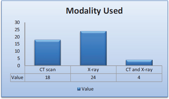
Figure 1. Types of imaging methods used in literature
3.5. Exploration of the Usage of Types of Data

Figure 2. Exploration of the usage of data type
4. Discussion
The global outbreak of COVID-19 has made medical imaging a crucial tool in the fight against the disease. Healthcare professionals rely on X-ray or CT images, along with PCR results, to accurately detect the virus. However, PCR results can sometimes be misleading, indicating other lung diseases such as pulmonary tuberculosis instead of COVID-19. This review paper provides a comprehensive analysis of previous studies on the diagnosis of COVID-19 using deep learning (DL) networks. The paper presents public databases available for COVID-19 diagnosis and prediction, as well as state-of-the-art DL techniques for diagnosis, segmentation, and forecasting of the disease spread. Table 1 in this paper outlines the DL techniques used for diagnosis and detection, respectively. The authors believe that with more public databases, researchers can develop more accurate DL models to detect and predict COVID-19. The features extracted from machine learning (ML) and DL models can also be used to improve accuracy in COVID-19 diagnosis.
Table 1. Classification of related work
Sr. No. |
Reference |
Aim |
Methodology |
Dataset used |
No of Images |
Imaging type |
Result |
1 |
COVID-19 pneumonia detection |
convolutional neural network (CNN) architecture and a transfer learning |
Open-source dataset from GitHub |
1,925 |
CT scans |
Accuracy: 93.5 %, Sensitivity: 92.9 %, Specificity: 93.8 % |
|
2 |
COVID-19 detection |
CNN |
NA |
NA |
Chest X-ray, CT Scan |
NA |
|
3 |
COVID-19 detection |
deep learning |
Public |
1,200 CT scans |
CT scans |
Accuracy:98.3 %, Sensitivity:97.1 %, Specificity:99.1 % |
|
4 |
COVID-19 detection |
VGG-19, ResNet-50, DenseNet-121 |
COVIDx dataset |
13,975 |
Chest X-ray |
Accuracy: 93.93 %. Sensitivity: 94.44 % Specificity:93.22 %, |
|
6 |
Detection. |
A literature review was conducted. |
Multiple datasets were used. |
-- |
Chest X-ray and CT scans. |
-- |
|
7 |
Detection |
hybrid deep neural network: (CNN)+ multilayer perceptron (MLP) network |
Public |
2000 images |
X-ray |
Accuracy: 94.8 % |
|
8 |
COVID-19 detection. |
ensemble + support vector machine (SVM) model |
Public dataset. |
330 |
Lung CT scans |
-- |
|
9 |
Detection |
machine learning |
Public datasets |
-- |
CTscans. |
Accuracy: 97.92 % |
|
10 |
COVID-19 detection |
VGG16 and ResNet50 deep learning algorithms |
Public dataset |
-- |
X-ray |
Accuracy: 96 % F1 score:0.95. |
|
11 |
COVID-19 detection |
QuNet |
Publicly available dataset |
-- |
X-ray |
Accuracy: 99 %, Precision:0.99, Recall:0.99, And F1 Score: 0.99. |
|
12 |
COVID-19 detection. |
CNN, VGG16 model. |
Public dataset |
1,200 |
Chest X-ray |
Accuracy:98 %, Precision:0.98, Recall: 0.98, F1 Score:0.98. |
|
13 |
COVID-19 detection |
VGG19, ResNet50, InceptionV |
Public dataset |
6,334 |
X-ray |
Accuracy: 97.24 %, Precision: 0.976, Recall: 0.972, F1score:0.974. |
|
14 |
Covid-19 detection. |
deep learning |
Public dataset |
2400 |
X-ray |
Accuracy:96.1 %, Precision:96.4 % Recall: 97.4 %, F1 Score: 96.9 %. |
|
15 |
Detection |
CNN |
Public dataset |
NA. |
CT scans |
Accuracy:96.5 % Sensitivity: 93.5 %, Specificity:98.5 %, F1 Score: 0.94. |
|
16 |
COVID-19 diagnosis |
machine learning, SVM, CNN |
Public dataset. |
498 |
Chest CT scan |
Accuracy: 94.18 %, Sensitivity:94.10 %, Specificity:94.26 %. SVM Accuracy: 90.16 %, CNN Accuracy: 93.18 %. |
|
17 |
COVID-19 diagnosis |
deep learning |
Private |
5,618 |
CT scans |
Sensitivity: 83.5 % Specificity: 98.5 %. |
|
18 |
COVID-19 detection |
deep learning |
Private |
900 |
X-ray |
Accuracy:96.46, Sensitivity:96.11 %, Specificity:96.81 %, AUC :0.9901. |
|
19 |
COVID-19 detection |
LeNet-5 CNN architecture |
Private. |
618 |
CT scans |
Accuracy:96.11 %, Sensitivity:96.42 %, Specificity:95.79 % Auc-Roc: 0.99. |
|
20 |
COVID-19 detection |
Faster RCNN, YOLO3, and SSD |
Private (China) |
Not specified |
CT scan. |
-- |
|
21 |
COVID-19 diagnosis |
InceptionV3, CNN |
Public |
5000 |
Chest X-ray images |
Accuracy:96.2 %,Sensitivity:96.0 % Specificity:96.4 %.. |
|
22 |
COVID-19 diagnosis |
Deep learning |
Private |
120 |
X-ray images |
Accuracy:95.5 % Sensitivity: 95.2 % Specificity:95.7 %. |
|
23 |
detection |
deep learning |
COVID-19 image database |
930 images |
X-ray |
Accuracy:98.7 % |
|
24 |
COVID-19 detection |
VGG-19, ResNet-50, InceptionV3 |
Public dataset |
1898 |
Chest X-ray images |
Accuracy:98.36 % Sensitivity:97.05 % Specificity:99.52 %. |
|
25 |
COVID-19 diagnosis |
machine learning CNN +SVM |
Public |
700 |
Chest X-ray images |
Accuracy:97.6 % Sensitivity:97 % Specificity:98 %. |
|
26 |
COVID-19 detection |
deep learning |
Private |
349 |
CT-scan images |
Accuracy:97.71 % Sensitivity: 98 % Specificity:97.5 %. |
|
27 |
COVID-19 detection |
Inception-V3 Softmax |
Public |
1560 |
CT scan |
Accuracy:98.5 % |
|
28 |
COVID-19 detection |
ResNet50 |
Public |
3616 |
X-ray |
Accuracy:98.4 % AUC Score: 0.99 |
|
29 |
COVID-19 detection |
VGG16 |
Public |
248 |
CT |
Accuracy: 94.7 % |
|
30 |
COVID-19 detection |
AlexNet |
Public |
531 |
CT |
Accuracy:91.91 % |
|
31 |
Feature detection |
nCoV |
Private |
1014 |
CT Images |
Ground-Glass Opacities (56.4 %) Bilateral Involvement (51.8 %). |
|
32 |
COVID-19 detection |
VGG16, ResNet50, InceptionV3 |
Public |
1764 |
X-ray and CT scan |
Accuracy:94.3 %, Sensitivity:95.5 %, Specificity:92.9 % |
|
33 |
COVID-19 detection |
VGG19 |
Public |
618 |
CT Images |
Accuracy:93.16 %, Sensitivity:91.98 %, Specificity: 94.41 %. |
|
34 |
Detection |
CVDNet |
Public and Private |
595 |
X-ray |
Public Dataset: Accuracy:93.9 % F1-Score:0.94 Private Datset: Accuracy:97.7 % F1-Score: 0.98 |
|
35 |
COVID-19 and pneumonia detection |
multi-dilation convolutional neural network |
Public |
11,960 |
X-ray |
-- |
|
36 |
Detection |
Hybrid model(VGG-16 + Inception V3 + ResNet-50) SVM classifier |
Private |
1,109 |
X-ray |
-- |
|
37 |
COVID-19 detection |
CNN, SVM |
Public |
812 |
CT scans |
Accuracy: 94.9 %, Sensitivity:92.3 %, Specificity: 96.4 %. |
|
39 |
Detection and classification |
CovXNet. |
Public |
9486 |
X-ray |
Accuracy:97.70 %Sensitivity:97.05 %, Specificity:97.79 %, F1-Score: 97.27 % |
|
40 |
detection and diagnosis |
DNN CoroNet |
Public |
-- |
X-ray |
Accuracy:96.78 % |
|
41 |
Detection |
convolutional neural network (CNN) |
Public |
-- |
CT scan |
Accuracy:95.25 % |
|
42 |
detection |
machine learning. |
Public. |
-- |
X-ray and CT |
Accuracy:96.5 %. |
|
44 |
Detection Networks. |
CapsNet |
Public |
28,968 |
X-ray |
Accuracy:98.86 % Sensitivity:98.81 %. |
|
45 |
Lesion segmentation |
U-net |
ISBI |
-- |
Medical |
-- |
|
46 |
detection |
deep learning. |
Public |
-- |
Chest X-ray images |
-- |
The deep learning models have achieved greater accuracy in detection as well as diagnosis. A deep learning model with a public dataset from CT scan images achieved an accuracy of 98.3 %, a sensitivity of 97.1 % and a specificity99.1 % (Bekhet et al., 2021). According to the literature, the accuracy of the deep learning model varies from 91 % to 98 %, depending on algorithm used.
At the initial stage, Covid-19 and seasonal flue share the same symptoms, so it is very difficult to differentiate them. According to the literature, several studies have used Al, machine learning and deep learning models to differentiate between Covid-19, bacterial and viral pneumonia. Some of the research studies have used CNN as base technology and some have used train test split methods.
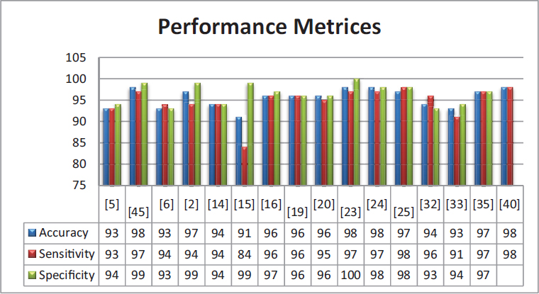
Figure 3. Performance metrics
Future studies in this area can be extended by using better deep learning models and larger datasets. Most of the researchers have used publicly available dataset from China and some of them have used private datasets. Thus, there is a greater scope to explore similarity patterns between Chinese datasets and local regional datasets. Also, this research has been conducted from the point of view of disease detection and diagnosis, however, it would also be important to research conduct it from the perspective of patient management, epidemic forecasting, and sustainable development.
5. Research Gap Analysis
Many studies on AI-based COVID-19 detection rely on limited and heterogeneous datasets. Addressing this gap involves the creation of standardized, large-scale, and diverse datasets that encompass various COVID-19 manifestations, age groups, and comorbidities. A well-annotated benchmark dataset will facilitate the development of more robust and generalizable AI models. Despite the remarkable performance of AI models, there is often a lack of transparency and interpretability in their decision-making processes. Research is needed to develop explainable AI models that can provide insights into the features and patterns that influence their predictions, improving trust and aiding clinical decision-making. Many AI-based COVID-19 detection models have been developed in controlled research settings. The gap lies in their real-world clinical validation and practical implementation. Future research should focus on validating the performance of AI models in clinical environments and addressing issues related to integration with existing healthcare systems.
Chest CT scans are valuable, but integrating them with other data modalities (e.g., clinical, genomic, or laboratory data) can enhance the accuracy and reliability of COVID-19 detection. Research should explore methods for effective multi-modal data fusion and analysis. Most studies focus on the detection of COVID-19 at a single point in time. Longitudinal studies, tracking changes in chest CT scans over time, can provide valuable insights into disease progression, treatment response, and recovery patterns, which are currently underexplored. Many AI models developed for COVID-19 detection are trained on data from specific populations, which can limit their generalizability. Research should aim to create models that are robust across diverse demographics, ethnicities, and healthcare systems.
Beyond mere detection, research can focus on developing AI systems that can aid healthcare providers in clinical decision-making. This involves not only accurate diagnosis but also prognosis, treatment recommendations, and risk stratification. Research on AI-based COVID-19 detection often focuses on well-resourced healthcare settings. Addressing the gap in resource-limited settings is crucial, where access to advanced diagnostic tools and expert radiologists may be limited. Exploring how AI can empower patients by providing them with tools for self-monitoring and early symptom detection is an emerging area of research. It encompasses mobile applications, wearable devices, and telemedicine solutions.
6. Future Scope
The approach to the detection of Covid-19 can be further modified by using an ensemble approach which can include more datasets, a variety of features and the fusion of various classifiers. The work can be enhanced by incorporating additional data modalities, such as clinical data and other medical imaging modalities such as MRI and X-ray, which could provide complementary information to further improve diagnosis and decision-making. The work related to COVID-19 early detection can be extended to real-world scenarios, ultimately benefiting healthcare by improving the accuracy of disease detection, treatment planning, and patient care.
The use of integrated AI and machine learning techniques will enhance both the accuracy and efficiency of existing approaches. Given that data availability is a critical factor in biomedical imaging, more diverse datasets are key to improving the robustness of the algorithm. After analyzing the recent literature on COVID-19 detection, it has been noticed that the early-stage detection of COVID-19 plays a crucial role in the healthcare area. For early detection, clinical and medical features as well as imaging features can be integrated.
References
Abdulateef, A. A., Mohammed, A. H., & Abdulateef, I. A. (2021). The Avoidance and Detection Function of Artificial Intelligence in Covid-19. HORA 2021 - 3rd International Congress on Human-Computer Interaction, Optimization and Robotic Applications, Proceedings. https://doi.org/10.1109/HORA52670.2021.9461280
Adeshina, S. A. (2022). A deep Learning Based methodology.
Agrawal, T., & Choudhary, P. (2022). FocusCovid: automated COVID-19 detection using deep learning with chest X-ray images. Evolving Systems, 13(4), 519–533. https://doi.org/10.1007/s12530-021-09385-2
Akter, S., Shamrat, F. M. J. M., Chakraborty, S., Karim, A., & Azam, S. (2021). Covid-19 detection using deep learning algorithm on chest X-ray images. Biology, 10(11). https://doi.org/10.3390/biology10111174
Alasasfeh, H. O., Alomari, T., & Ibbini, M. S. (2021). Deep Learning Approach for COVID-19 Detection Based on X-Ray Images. 18th IEEE International Multi-Conference on Systems, Signals and Devices, SSD 2021, January, 601–606. https://doi.org/10.1109/SSD52085.2021.9429383
Alazab, M., Awajan, A., Mesleh, A., Abraham, A., Jatana, V., & Alhyari, S. (2020). COVID-19 Prediction and Detection Using Deep Learning. In International Journal of Computer Information Systems and Industrial Management Applications (Vol. 12). www.mirlabs.net/ijcisim/index.html
Alqudah, A. M., Qazan, S., & Alqudah, A. (2020). Automated Systems for Detection of COVID-19 Using Chest X-ray Images and Lightweight Convolutional Neural Networks. Emergency Radiology, 4(1), 54–67. https://www.researchsquare.com/article/rs-24305/latest.pdf
Ardakani, A. A., Kanafi, A. R., Acharya, U. R., Khadem, N., & Mohammadi, A. (2020). Application of deep learning technique to manage COVID-19 in routine clinical practice using CT images: Results of 10 convolutional neural networks. Computers in Biology and Medicine, 121. https://doi.org/10.1016/j.compbiomed.2020.103795
Asghar, U., Arif, M., Ejaz, K., Vicoveanu, D., Izdrui, D., & Geman, O. (2022). An Improved COVID-19 Detection using GAN-Based Data Augmentation and Novel QuNet-Based Classification. BioMed Research International, 2022. https://doi.org/10.1155/2022/8925930
Badrinarayanan, V., Kendall, A., & Cipolla, R. (2017). \href{https://arxiv.org/pdf/1511.00561.pdf}{Segnet: A deep convolutional encoder-decoder architecture for image segmentation}. IEEE Transactions on Pattern Analysis and Machine Intelligence, 39(12), 2481–2495. https://arxiv.org/pdf/1511.00561.pdf
Bekhet, S., Alkinani, M. H., Tabares-Soto, R., & Hassaballah, M. (2021). An efficient method for covid-19 detection using light weight convolutional neural network. Computers, Materials and Continua, 69(2), 2475–2491. https://doi.org/10.32604/cmc.2021.018514
Bekhet, S., Hassaballah, M., Kenk, M. A., & Hameed, M. A. (2020). An Artificial Intelligence Based Technique for COVID-19 Diagnosis from Chest X-Ray. 2nd Novel Intelligent and Leading Emerging Sciences Conference, NILES 2020, 191–195. https://doi.org/10.1109/NILES50944.2020.9257930
Chung, M., Bernheim, A., Mei, X., Zhang, N., Huang, M., Zeng, X., Cui, J., Xu, W., Yang, Y., Fayad, Z. A., Jacobi, A., Li, K., Li, S., & Shan, H. (2020). CT imaging features of 2019 novel coronavirus (2019-NCoV). Radiology, 295(1), 202–207. https://doi.org/10.1148/radiol.2020200230
Das, A. K., Ghosh, S., Thunder, S., Dutta, R., Agarwal, S., & Chakrabarti, A. (2021). Automatic COVID-19 detection from X-ray images using ensemble learning with convolutional neural network. Pattern Analysis and Applications, 24(3), 1111–1124. https://doi.org/10.1007/s10044-021-00970-4
Das, D., Biswas, S. K., & Bandyopadhyay, S. (2022). Perspective of AI system for COVID-19 detection using chest images: a review. Multimedia Tools and Applications, 81(15), 21471–21501. https://doi.org/10.1007/s11042-022-11913-4
Dhebe, R., Jagtap, V., Munde, P., & Salian, P. S. (2019). Covid-19 Detection using X-ray. 3307, 147–153.
Duong, L. T., Nguyen, P. T., Iovino, L., Flammini, M., & Linh, L. T. (2020). Deep Learning for Automated Recognition of Covid-19 from Chest X-ray Images. MedRxiv, August, 2020.08.13.20173997. https://doi.org/10.1101/2020.08.13.20173997 %0Ahttps://www.medrxiv.org/content/10.1101/2020.08.13.20173997v1.abstract
Feng, K., He, F., Steinmann, J., & Demirkiran, I. (2021). Deep-learning based approach to identify covid-19. Conference Proceedings - IEEE SOUTHEASTCON, 2021-March, 17–20. https://doi.org/10.1109/SoutheastCon45413.2021.9401826
Foysal Haque, K., Farhan Haque, F., Gandy, L., & Abdelgawad, A. (2020). Automatic Detection of COVID-19 from Chest X-ray Images with Convolutional Neural Networks. Proceedings - 2020 International Conference on Computing, Electronics and Communications Engineering, ICCECE 2020, 125–130. https://doi.org/10.1109/iCCECE49321.2020.9231235
Halder, A., & Datta, B. (2021). COVID-19 detection from lung CT-scan images using transfer learning approach. Machine Learning: Science and Technology, 2(4). https://doi.org/10.1088/2632-2153/abf22c
Horry, M. J., Chakraborty, S., Paul, M., Ulhaq, A., Pradhan, B., Saha, M., & Shukla, N. (2020). COVID-19 Detection through Transfer Learning Using Multimodal Imaging Data. IEEE Access, 8, 149808–149824. https://doi.org/10.1109/ACCESS.2020.3016780
Islam, M. R., & Matin, A. (2020). Detection of COVID 19 from CT Image by the Novel LeNet-5 CNN Architecture. ICCIT 2020 - 23rd International Conference on Computer and Information Technology, Proceedings, 19–21. https://doi.org/10.1109/ICCIT51783.2020.9392723
Kaheel, H., Hussein, A., & Chehab, A. (2021). AI-Based Image Processing for COVID-19 Detection in Chest CT Scan Images. Frontiers in Communications and Networks, 2(August), 1–12. https://doi.org/10.3389/frcmn.2021.645040
Kassania, S. H., Kassanib, P. H., Wesolowskic, M. J., Schneidera, K. A., & Detersa, R. (2021). Automatic Detection of Coronavirus Disease (COVID-19) in X-ray and CT Images: A Machine Learning Based Approach. Biocybernetics and Biomedical Engineering, 41(3), 867–879. https://doi.org/10.1016/j.bbe.2021.05.013
Khan, A. I., Shah, J. L., & Bhat, M. M. (2020). CoroNet: A deep neural network for detection and diagnosis of COVID-19 from chest x-ray images. Computer Methods and Programs in Biomedicine, 196. https://doi.org/10.1016/j.cmpb.2020.105581
Khan, A., Younis, S., & Algethami, H. (2021). Covid-19 Identification using deep neural networks. 2021 International Conference of Women in Data Science at Taif University, WiDSTaif 2021. https://doi.org/10.1109/WIDSTAIF52235.2021.9430219
Kieu, S. T. H., Bade, A., Hijazi, M. H. A., & Kolivand, H. (2021). COVID-19 Detection Using Integration of Deep Learning Classifiers and Contrast-Enhanced Canny Edge Detected X-Ray Images. IT Professional, 23(4), 51–56. https://doi.org/10.1109/MITP.2021.3052205
Laguarta, J., Hueto, F., & Subirana, B. (2020). COVID-19 Artificial Intelligence Diagnosis Using only Cough Recordings. IEEE Open Journal of Engineering in Medicine and Biology, 1, 275–281. https://doi.org/10.1109/OJEMB.2020.3026928
Liu, J., Zhang, Z., Zu, L., Wang, H., & Zhong, Y. (2020). Intelligent Detection for CT Image of COVID-19 using Deep Learning. Proceedings - 2020 13th International Congress on Image and Signal Processing, BioMedical Engineering and Informatics, CISP-BMEI 2020, 76–81. https://doi.org/10.1109/CISP-BMEI51763.2020.9263690
Mahmud, T., Rahman, M. A., & Fattah, S. A. (2020). CovXNet: A multi-dilation convolutional neural network for automatic COVID-19 and other pneumonia detection from chest X-ray images with transferable multi-receptive feature optimization. Computers in Biology and Medicine, 122. https://doi.org/10.1016/j.compbiomed.2020.103869
Meng, Z., Wang, M., Song, H., Guo, S., Zhou, Y. Y. Y., Li, W., Zhou, Y. Y. Y., Li, M., Song, X., Zhou, Y. Y. Y., Li, Q., Lu, X., & Ying, B. (2020). Development and utilization of an intelligent application for aiding COVID-19 diagnosis. MedRxiv, 37, https://doi.org/2020.03.18.20035816. https://www.medrxiv.org/content/10.1101/2020.03.18.20035816v1
Mishra, M., Parashar, V., & Shimpi, R. (2020). Development and evaluation of an AI System for early detection of Covid-19 pneumonia using X-ray (Student Consortium). Proceedings - 2020 IEEE 6th International Conference on Multimedia Big Data, BigMM 2020, 292–296. https://doi.org/10.1109/BigMM50055.2020.00051
Mishra, N. K., Singh, P., & Joshi, S. D. (2021). Automated detection of COVID-19 from CT scan using convolutional neural network. Biocybernetics and Biomedical Engineering, 41(2), 572–588. https://doi.org/10.1016/j.bbe.2021.04.006
Nair, R., Vishwakarma, S., Soni, M., Patel, T., & Joshi, S. (2022). Detection of COVID-19 cases through X-ray images using hybrid deep neural network. World Journal of Engineering, 19(1), 33–39. https://doi.org/10.1108/WJE-10-2020-0529
Ouchicha, C., Ammor, O., & Meknassi, M. (2020). CVDNet: A novel deep learning architecture for detection of coronavirus (Covid-19) from chest x-ray images. Chaos, Solitons and Fractals, 140. https://doi.org/10.1016/j.chaos.2020.110245
Ozturk, T., Talo, M., Azra, E., Baran, U., & Yildirim, O. (2020). Since January 2020 Elsevier has created a COVID-19 resource centre with free information in English and Mandarin on the novel coronavirus COVID- 19 . The COVID-19 resource centre is hosted on Elsevier Connect , the company ’ s public news and information. Computers in Biology and Medicine, January.
Patgar, C. C., Patil, D. D., Rahate, S. H., & Randive, S. (2022). Covid-19 Detection using Deep Learning. 2022 International Conference on Signal and Information Processing, IConSIP 2022. https://doi.org/10.1109/ICoNSIP49665.2022.10007465
Pont-Tuset, J., & Marques, F. (2016). Supervised Evaluation of Image Segmentation and Object Proposal Techniques. IEEE Transactions on Pattern Analysis and Machine Intelligence, 38(7), 1465–1478. https://doi.org/10.1109/TPAMI.2015.2481406
Sangidong, J. C., Purnomo, H. D., & Santoso, F. Y. (2021). Application of Deep Learning for Early Detection of COVID-19 Using CT-Scan Images. 3rd 2021 East Indonesia Conference on Computer and Information Technology, EIConCIT 2021, 61–65. https://doi.org/10.1109/EIConCIT50028.2021.9431887
Shadin, N. S., Sanjana, S., & Lisa, N. J. (2021). COVID-19 Diagnosis from Chest X-ray Images Using Convolutional Neural Network(CNN) and InceptionV3. 2021 International Conference on Information Technology, ICIT 2021 - Proceedings 3 (September 2012), 799–804. https://doi.org/10.1109/ICIT52682.2021.9491752
Shah, F. M., Joy, S. K. S., Ahmed, F., Hossain, T., Humaira, M., Ami, A. S., Paul, S., Jim, M. A. R. K., & Ahmed, S. (2021). A Comprehensive Survey of COVID-19 Detection Using Medical Images. SN Computer Science, 2(6), 1–22. https://doi.org/10.1007/s42979-021-00823-1
Sharma, S., & Tiwari, S. (2021). COVID-19 Diagnosis using X-Ray Images and Deep learning. Proceedings - International Conference on Artificial Intelligence and Smart Systems, ICAIS 2021, 344–349. https://doi.org/10.1109/ICAIS50930.2021.9395851
Shrivastava, P., Singh, A., Agarwal, S., Tekchandani, H., & Verma, S. (2021). Covid detection in CT and X-Ray images using Ensemble Learning. Proceedings - 5th International Conference on Computing Methodologies and Communication, ICCMC 2021, Iccmc, 1085–1090. https://doi.org/10.1109/ICCMC51019.2021.9418308
Singh, M., Bansal, S., Ahuja, S., Dubey, R. K., Panigrahi, B. K., & Dey, N. (2021). Transfer learning–based ensemble support vector machine model for automated COVID-19 detection using lung computerized tomography scan data. Medical and Biological Engineering and Computing, 59(4), 825–839. https://doi.org/10.1007/s11517-020-02299-2
Toraman, S., Alakus, T. B., & Turkoglu, I. (2020). Convolutional capsnet: A novel artificial neural network approach to detect COVID-19 disease from X-ray images using capsule networks. Chaos, Solitons and Fractals, 140. https://doi.org/10.1016/j.chaos.2020.110122
Wang, T., Zhao, Y., Zhu, L., Liu, G., Ma, Z., & Zheng, J. (2020). Lung CT image aided detection COVID-19 based on Alexnet network. Proceedings - 2020 5th International Conference on Communication, Image and Signal Processing, CCISP 2020, 199–203. https://doi.org/10.1109/CCISP51026.2020.9273512
Weng, W., & Zhu, X. (2021). INet: Convolutional Networks for Biomedical Image Segmentation. IEEE Access, 9, 16591–16603. https://doi.org/10.1109/ACCESS.2021.3053408
Yang, D., Martinez, C., Visuña, L., Khandhar, H., Bhatt, C., & Carretero, J. (2021). Detection and analysis of COVID-19 in medical images using deep learning techniques. Scientific Reports, 11(1), 1–13. https://doi.org/10.1038/s41598-021-99015-3
Zhao, W., Jiang, W., & Qiu, X. (2021). Deep learning for COVID-19 detection based on CT images. Scientific Reports, 11(1), 1–12. https://doi.org/10.1038/s41598-021-93832-2